
About this note
A plain-language summary of the 8 publications listed under this topic on Academic Publications (Topic-wise). Full citations are under References.
Why this matters
The eye is unusually exposed to heat. Parts of it, such as the cornea and lens, have no blood flow to carry heat away, and it has no thermal sensors or protective reflex beyond blinking (Narasimhan, 2017). Yet lasers are routinely aimed into it. In pan-retinal photocoagulation (PRP), used for diabetic retinopathy and other retinal diseases, some 700 to 1,500 small laser spots are burned into the retina (Narasimhan and Jha, 2011). The target tissue needs only about 60 °C to coagulate, but overheating can damage healthy tissue next to it (Narasimhan et al., 2010). Temperatures inside a living human eye cannot be measured directly during surgery, so numerical simulation is one of the few ways to see what the laser actually does and to suggest safer settings.
The heat is absorbed mainly in the retinal pigmented epithelium (RPE), a thin pigmented layer at the back of the retina. Behind it lies the choroid, a layer rich in blood vessels that was long assumed to be the eye’s main cooling system. Much of this research line, carried out mostly with Kaushal Kumar Jha during his doctoral work, tested that assumption and looked for ways to keep the RPE near 60 °C without collateral damage.
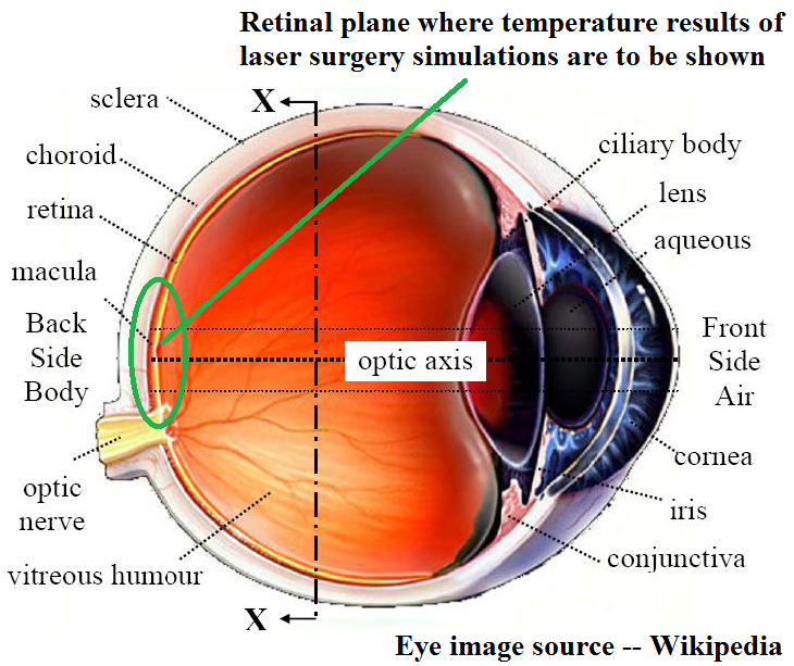
Figure 1. Parts of the human eye. The laser enters through the cornea and lens at the front and is absorbed at the back, in the RPE and the pigmented choroid next to it. The green circle marks the retinal region where the simulated temperatures are reported. Eye image source: Wikipedia.
The pigmented layers turn the absorbed light into heat, and the local temperature climbs well above the body core temperature of 37 °C. For treatment, each spot should be heated just enough to coagulate, to about 60 °C and no more, because overheating disrupts the cells of the healthy tissue around it. Using the specifications of an actual surgery, with a 0.2 W laser and a 500 µm spot, our simulations predicted peak retinal temperatures of 104 °C after a 100 ms shot and 172 °C if the heating were held to steady state (Narasimhan et al., 2010).
What we did
All the models solve the Pennes bio-heat equation, which adds a blood perfusion term (the cooling by blood flowing through tissue) to ordinary heat conduction, using the finite volume method on a realistic eye geometry with properties for each tissue layer.
A single laser spot in two dimensions
With ophthalmologist Lingam Gopal of Sankara Nethralaya, we first built and validated a two-dimensional model of the whole eye and followed the temperature during a 100 ms laser exposure (Narasimhan et al., 2010). Choroidal blood flow was represented both by a perfusion term and by a convection condition at the sclera (the eye’s white outer wall). A follow-up study added the effect of choroidal pigmentation, the pigment in the choroid that also absorbs some laser energy, and extended the model to a seven-spot circular pattern in three dimensions (Narasimhan and Jha, 2012).
Many spots in three dimensions
Real surgery fires many spots, so we built a truncated three-dimensional model of the retinal region and fired a 3 × 3 square array of 0.2 W spots one after another, varying the centre-to-centre spacing between spots (Narasimhan and Jha, 2010). We then compared square arrays with circular arrays of six spots around a centre spot (Narasimhan and Jha, 2011), and sequential firing with simultaneous firing of all spots (Jha and Narasimhan, 2011). In each case we tried two remedies for overheating: pulsing the laser on and off after the target first reaches 60 °C, and simply reducing the laser power.
The laser settings in these simulations were taken from the Pascal pattern-scanning laser, which places a whole array of spots in one go (about the Pascal laser). This video of our simulations shows the temperature on the retina rising and spreading as the spots are fired one after another.
To compare with the real thing, here is a video of an actual retinal laser surgery, courtesy of Dr. Lehr, MD.
Fluid flow inside the eye during thermotherapy
Transpupillary thermotherapy (TTT) uses a long, low-intensity infrared laser exposure over a large spot. After some retinal operations, or with age, the gel-like vitreous humour filling the eye is replaced by a watery liquid that can move. With Sundarraj C., we solved the flow and energy equations together to see whether natural convection (flow driven by warm fluid rising and cool fluid sinking) in this liquid changes retinal heating during TTT (Narasimhan and Sundarraj, 2013). The flow side of this story is told in Bio-Fluid Dynamics of Human Eye.
Reviews
Two book chapters place this work in context. The first, again with Lingam Gopal, reviews laser-tissue interaction, the history of eye heat transfer models and our methods for limiting thermal damage (Narasimhan and Gopal, 2012). The second, written for the Handbook of Thermal Science and Engineering, surveys heat and mass transfer in the eye more broadly: evaporation of the tear film, laser treatments from cornea to retina, cryotherapy, drug delivery to the retina, and the combination of heating and drug transport through the vitreous (Narasimhan, 2017).
What we found
Choroidal blood flow does not cool the retina during a laser shot. At steady state, choroidal blood flow does lower the RPE temperature. But during a 100 ms exposure the RPE reached about 101 to 105 °C regardless of choroidal flow, and the scleral boundary condition made no difference either (Narasimhan et al., 2010). The reason is a mismatch of time scales: heat needs much longer than 100 ms to diffuse from the retina to the choroid. The cornea stayed at about 43 °C, too low for photocoagulation there. Later work confirmed that perfusion cooling is negligible in multi-spot simulations too, whereas choroidal pigmentation does matter, lowering the peak temperature from 103 °C to 94 °C at the same 0.2 W power because more pigmented tissue shares the absorbed energy (Narasimhan and Jha, 2012).
The laser overshoots, and pulsing helps. The RPE needs only about 20 ms to reach 60 °C, so the rest of a 100 ms exposure mostly heats surrounding tissue (Narasimhan et al., 2010). Switching the laser between high and low levels kept the RPE near the target. In the array simulations, pulsing brought the average peak temperature of the spots down to about 70 °C, and reducing the power to 0.072 W held it near 60 °C (Narasimhan and Jha, 2011; Narasimhan and Gopal, 2012). Pulsing to keep the peak within 10% of 60 °C was proposed as a way to reduce long-term thermal damage, whatever the spot spacing (Narasimhan and Jha, 2010).
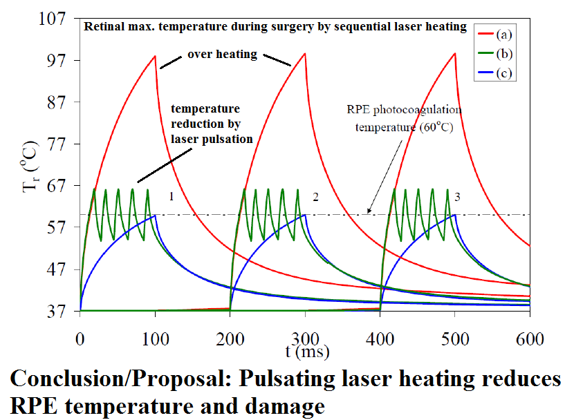
Figure 2. Peak retinal temperature as three spots are fired one after another, 200 ms apart, each for 100 ms. (a) At a steady 0.2 W the RPE overshoots to nearly 100 °C on every shot. (b) Pulsing the laser between a high and a low level after the first 60 °C holds it within a few degrees of the target. (c) At reduced power the spot climbs to 60 °C just as the shot ends.
Spots need room. When spots were 0.375 mm apart or closer, untreated retina between them rose to 60 °C, and each successive spot in a row ran hotter, by up to 10 °C within 400 ms (Narasimhan and Jha, 2010). At about 0.6 mm or more, heat diffused away enough for the retina to fall back close to body temperature between shots. Square and circular arrays behaved almost identically, with peak temperatures differing by less than 0.6 °C, and about 0.625 mm emerged as a suitable spacing (Narasimhan and Jha, 2011). Comparing firing modes, sequential firing ran about 2 °C hotter because each spot was preheated by its neighbours but cooled faster, while simultaneous firing is many times quicker, which matters when 1,500 to 1,600 spots must be placed; with spots at least 0.75 mm apart the two modes cooled alike (Jha and Narasimhan, 2011).
A moving vitreous changes the picture. In TTT, natural convection in a liquefied vitreous lowered the peak retinal temperature by 15 K at steady state and 12.5 K after 60 s, compared with a stationary vitreous, while choroidal perfusion lowered it by 6 K and 1.5 K (Narasimhan and Sundarraj, 2013). The flow also carried heat to retina next to the laser spot, warming it by about 2 K, so treating the vitreous as stagnant can understate thermal damage there.
The reviews close on the same caution: these are simulations, and experimental measurements in functioning human eyes are still needed, which only interdisciplinary teams of doctors, engineers and biologists can supply (Narasimhan and Gopal, 2012; Narasimhan, 2017). A related study showed that for millisecond laser pulses, the finite speed of heat conduction in tissue may also matter; see Non-Fourier Bio-heat Transfer.
Related
References
- Narasimhan, A., (2017), “Heat and Mass Transfer Processes in the Eye,” pp. 1-35, in Handbook of Thermal Science and Engineering, ed. F. A. Kulacki, Springer International Publishing. [DOI: 10.1007/978-3-319-32003-8_72-1 | Abstract]
- Narasimhan, A. and Sundarraj, C., (2013), “Effect of choroidal blood perfusion and natural convection in vitreous humor during transpupillary thermotherapy (TTT),” Int. J. Numer. Meth. Biomed. Engng., [DOI: 10.1002/cnm.2538 | Abstract]
- Narasimhan, A. and Gopal, L., (2012), “Modeling Retinal Laser Surgery in Human Eye,” Chapter 11, in Human Eye Imaging and Modeling (Eds. E. Y. K. Ng et al.), CRC Press, Taylor and Francis Group, NY. [ product link ].
- Narasimhan, A. and Jha, K. K., (2012), “Bio-Heat Transfer Simulation of Retinal Laser Irradiation,” Int. J. Numer. Meth. Biomed. Engng. [DOI: 10.1002/cnm.1489]
- Narasimhan, A. and Jha, K. K., (2011), “Bio-Heat Transfer Simulation of Square and Circular Array of Retinal Laser Irradiation,” Frontiers in Heat and Mass Transfer, **2, **033005 [DOI: http://dx.doi.org/10.5098/hmt.v2.3.3005 | Abstract | PDF Download]
- Jha, K. K. and Narasimhan, A., (2011), “Three-Dimensional Bio-Heat Transfer Simulation of Sequential and Simultaneous Retinal Laser Irradiation,” Int. J. Thermal Sci., 50(7), 1191-1198. [DOI:10.1016/j.ijthermalsci.2011.02.005]
- Narasimhan, A. and Jha, K. K., (2010), “Transient Simulation of Multi-spot Retinal Laser Irradiation using Bio-Heat Transfer Model”, Numerical Heat Transfer, Part A: Applications, 57(7), 520-536. [DOI:10.1080/10407781003684514 ]
- Narasimhan, A., Jha, K. K. and Gopal, L., (2010), “Transient Simulations of Heat Transfer in Human Eye undergoing Laser Surgery”, Int. J. Heat and Mass Transfer, 53 (1), 482-490. [DOI 10.1016/j.ijheatmasstransfer.2009.09.007].